 Lineora 3D Modelss


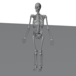
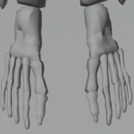

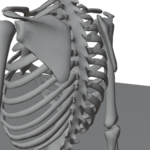
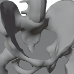
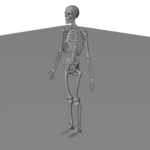
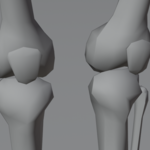
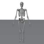
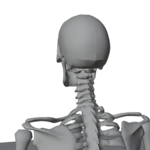
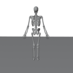
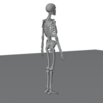
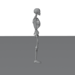
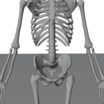
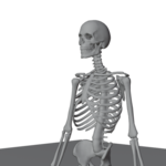
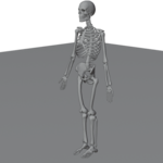
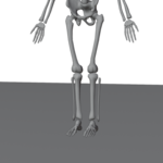
This realistic 3D illustration showcases the complete human skeletal system in an upright standing position, accurately representing the anatomical structure and alignment of bones throughout the body. The model clearly displays major skeletal components including the skull, rib cage, spine, pelvis, arms, hands, legs, and feet, making it an excellent visual reference for understanding human bone anatomy.
Rendered in a clean, neutral environment, the image is ideal for medical education, anatomy studies, biology lessons, healthcare presentations, orthopedic research, and digital learning materials. The simplified yet anatomically correct design allows students and professionals to easily identify bone placement, joint connections, and overall skeletal proportions.
This illustration is especially useful for medical students, anatomy instructors, physiotherapists, healthcare professionals, and content creators looking for a clear and accurate 3D representation of the human skeletal system.